tree in bud lesion
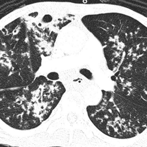
The tree-in-bud pattern is commonly seen at thin-section computed tomography CT of the lungs. Cavitary lesion with decreased lung parenchymal volume is seen at right upper lobe.
2
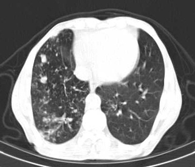
The small nodules represent lesions involving the small airways.

. At examination with CT centrilobular lesions nodules or branching linear structures 2-4 mm in diameter were most commonly seen n 39 95. Without an obvious mass although a small central lesion is not excluded. Based on lesion localization and presence or suspicion of a concomitant bronchial disease for cats in this sample authors propose that the CT tree-in-bud pattern described.
Cavitary lesion with decreased lung. Centrilobular nodules with a linear branching pattern are consistent with tree-in-bud appearance in a patient with endobronchial spreading of post-primary tuberculosis. It consists of small centrilobular nodules of soft-tissue attenuation connected to multiple branching linear structures of similar caliber that originate from a single stalk.
Usually somewhat nodular in appearance the tree-in-bud pattern is generally most pronounced in the lung periphery and associated with abnormalities of the larger airways. The tree-in-bud sign is a nonspecific imaging finding that implies impaction within bronchioles the smallest airway passages in the lung. 87 rows mid-bronchial lesion.
Overlap of the clusters with multiple tree-in-bud lesions demonstrating clearly the tree bronchiole and the bud alveolar ducts Fig. The associated central bronchi are impacted. 1 refers to a pattern seen on thin-section chest CT in which centrilobular bronchial dilatation and filling by mucus pus or fluid resembles a budding tree Fig.
The patient had an oesophageal lesion below the carina extending longitudinally 6 cm. The only noninflammatory cause of TIB opacities in our study was lymphoma which has previously been reported. In the 26 patients with follow-up most of these.
The tree-in-bud-pattern of images on thin-section lung CT is defined by centrilobular branching structures that resemble a budding tree. We investigated the pathological basis of the tree-in-bud lesion by reviewing the pathological specimens of bronchograms of normal lungs and contract radiographs of the post-mortem lungs manifesting active pulmonary tuberculosis. The tree-in-bud-pattern of images on thin-section lung CT is defined by centrilobular branching structures that resemble a budding tree.
My CT scan says defined streaky opacity with associated loss volume and clustered tree in bud nodules have developed in the anterior segment of the upper left lobe. Non-infectious causes of the tree-in-bud sign include diffuse panbronchiolitis cystic fibrosis immotile cilia syndrome and congenital immunodeficiency. We investigated the pathological basis of the tree-in-bud lesion by reviewing the pathological specimens of bronchograms of normal lungs and contract radiographs of the post-mortem lungs manifesting.
Our Radiology Information System was searched for the term tree-in-bud from January 1 2010 to December 31 2010 identifying 599 examinations. In one section the clustered small nodules occurred predominantly in the peripheral portion of the secondary pulmonary lobule which can easily be misinterpreted as perilobular lesions on CT 20. Originally reported in cases of endobronchial spread of Mycobacterium tuberculosis this.
Post-mortem radiograph of patient with active pulmonary tuberculosis demonstrating tree-in-bud lesion boxed area with smooth marginated bronchiole tree and distal clubbed end bud. In radiology the tree-in-bud sign is a finding on a CT scan that indicates some degree of airway obstruction. Usually somewhat nodular in appearance the tree-in-bud pattern is generally most pronounced in the lung periphery and associated with abnormalities of the.
Obstructive airway lesions and bronchiolitis obliterans organizing pneumonia. The impression at the end said a focus of bronchitis and. In humans a CT tree-in-bud pattern has been described as a characteristic of centrilobular bronchiolar dilation with bronchiolar plugging by mucus pus or fluid.
The tree-in-bud sign could be seen in various infectious diseases including endobronchial spread of tuberculosis bacterial viral parasitic and fungal infections involving the bronchioles. In one section the clustered small nodules occurred predominantly in the peripheral portion of the secondary pulmonary lobule which can easily be misinterpreted as perilobular lesions on CT 20. Bronchial wall thickening branching micronodules producing a.

Tree In Bud Pattern Pulmonary Tb Eurorad

Tree In Bud Pattern Pulmonary Tb Eurorad

Tree In Bud Sign Lung Radiology Reference Article Radiopaedia Org
2
2
View Of Tree In Bud The Southwest Respiratory And Critical Care Chronicles

Areas Showing A Mosaic Pattern Of Attenuation And Tree In Bud Opacities Download Scientific Diagram

Computed Tomography Of The Chest Showed Nodular Opacities With Tree In Download Scientific Diagram

Tree In Bud Sign Lung Radiology Reference Article Radiopaedia Org

Pdf Tree In Bud

Tree In Bud Pattern Pulmonary Tb Eurorad

Tree In Bud Pattern Pulmonary Tb Eurorad
Tree In Bud Pattern Pulmonary Tb Eurorad
Tree In Bud Pattern Pulmonary Tb Eurorad

Tree In Bud Sign Lung Radiology Reference Article Radiopaedia Org
Tree In Bud Appearance Endobronchial Spreading Of Pulmonary Tuberculosis Radiology Case Radiopaedia Org

Co Rads 2 With Tree In Bud Sign A 27 Year Old Male Attended The Download Scientific Diagram
View Of Tree In Bud The Southwest Respiratory And Critical Care Chronicles

Figure 3 From Tree In Bud Pattern Semantic Scholar